|
14-011h
|
|
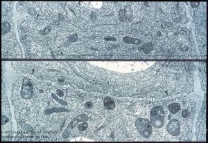
Two stages in sieve plate development: plasmodesmal stage and plasmodesmata partly lined with callose. (Nicotiana tabacum root tip)
|
copyright: Katherine Esau, BSA
license: http://images.botany.org/index.html#license |
Image
|
Cellular Communication Channels

|

|
|
14-012h
|
|
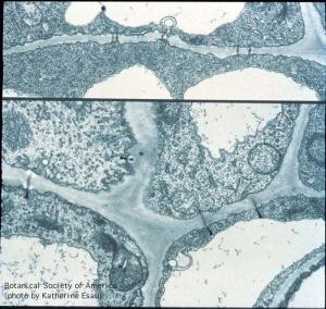
Young wall with plasmodesmata (top); sieve plate elements (left) and companion cells (right). Early stage of callose accumulation visible at ends of a plasmodesma, near differentiating pore site. (Echium angustifolium and E. sabulicola petioles)
|
copyright: Katherine Esau, BSA
license: http://images.botany.org/index.html#license |
Image
|
Cellular Communication Channels

|

|
|
14-013h
|
|
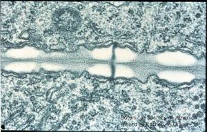
Further development of a pore site in longitudinal view. Callose nearly fused at middle lamella, plasmalemma and ER cisternae cover the callose, plasmodesma extends from ER to ER through future pore. (Echium angustifolium petiole)
|
copyright: Katherine Esau, BSA
license: http://images.botany.org/index.html#license |
Image
|
Cellular Communication Channels

|

|
|
14-014h
|
|
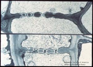
Continuous callose and plasmalemma in former plasmodesmal canal (above). Most callose was removed and open pores partly occluded with P-protein. (Echium angustifolium petioles)
|
copyright: Katherine Esau, BSA
license: http://images.botany.org/index.html#license |
Image
|
Cellular Communication Channels

|

|
|
14-015h
|
|
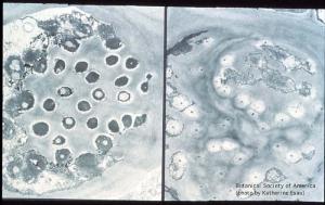
Left, plugs of callose, penetrated by plasmodesmata, fill the pores. Right, pores open, lined with thin layer of callose, and most are occluded with P-protein, some with starch grains. (Echium wildpreti petioles)
|
copyright: Katherine Esau, BSA
license: http://images.botany.org/index.html#license |
Image
|
Cellular Communication Channels

|

|
|
14-016h
|
|
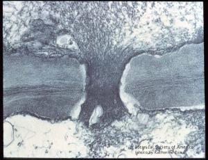
P-protein plug in pore in longitudinal section. Compaction of P-protein within pore especially through middle lamella region. Echium judaeum petiole
|
copyright: Katherine Esau, BSA
license: http://images.botany.org/index.html#license |
Image
|
Cellular Communication Channels

|

|
|
14-017h
|
|
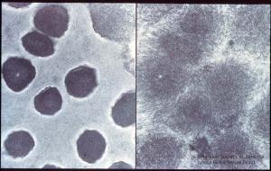
Cross sections through pores filled with P-protein and through masses of loose P-protein located at some distance from the sieve plate. Echium plantagineum petioles.
|
copyright: Katherine Esau, BSA
license: http://images.botany.org/index.html#license |
Image
|
Cellular Communication Channels

|

|
|
14-018h
|
|
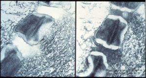
P-protein digestion with pepsin (left); control (right). Compacted P-protein within the sieve plate pore became digested. (Echium judaeum petiole)
|
copyright: Katherine Esau, BSA
license: http://images.botany.org/index.html#license |
Image
|
Cellular Communication Channels

|

|
|
14-019h
|
|
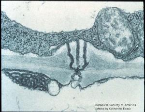
Longitudinal section of cell wall including a plasmodesma connecting a sieve element (below) and branched plasmodesma located on the companion cell side of the wall. (Echium rosulatum petiole)
|
copyright: Katherine Esau, BSA
license: http://images.botany.org/index.html#license |
Image
|
Cellular Communication Channels

|

|
|
14-020h
|
|
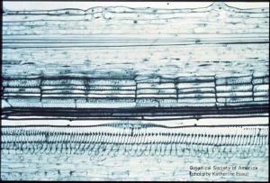
Primary xylem of Phaseolus vulgaris stem, with ring-like and helical secondary walls (close to bottom of slide). Secondary wall surrounds perforations at the two ends of an individual vessel member. Perforations are formed by localized removal of the primary wall.
|
copyright: Katherine Esau, BSA
license: http://images.botany.org/index.html#license |
Image
|
Cellular Communication Channels

|

|
|
14-021h
|
|
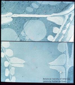
Removal of vessel end wall with middle part consisting of primary wall material. Around the end wall margin, secondary wall material protects unthickened primary wall, which becomes hydrolyzed and disappear. This stage is shown below: the rim now surrounds a perforation. (Phaseolus vulgaris stem)
|
copyright: Katherine Esau, BSA
license: http://images.botany.org/index.html#license |
Image
|
Cellular Communication Channels

|

|
|
14-022h
|
|
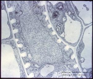
Unperforated vessel wall parts also undergo a change; primary wall not covered by secondary wall layers undergoes partial hydrolysis. Beta vulgaris leaf.
|
copyright: Katherine Esau, BSA
license: http://images.botany.org/index.html#license |
Image
|
Cellular Communication Channels

|

|
|
14-023h
|
|
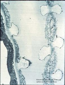
Primary wall between contiguous vessel members (right) is partially hydrolyzed between secondary thickening gyres. Noncellulosic wall components removed, cellulose forms a loose network. Hydrolysis affects primary wall adjacent to vessel member. (Capsella bursa pastoris leaf)
|
copyright: Katherine Esau, BSA
license: http://images.botany.org/index.html#license |
Image
|
Cellular Communication Channels

|

|
|
14-024h
|
|
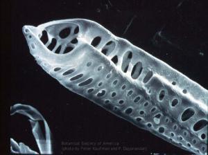
Scanning electron microscograph of vessel member from Pelargonium leaf with perforations and pits. Micrograph, courtesy of Professor Peter B. Kaufman and Dr. P Dayanandan, Dept. of Botany, Univ. of Michigan, Ann Arbor, MI.
|
copyright: Katherine Esau, BSA
license: http://images.botany.org/index.html#license |
Image
|
Cellular Communication Channels

|

|
|
15-001h
|
|
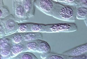
Prophase. Onion root tip cell [right] in mid-prophase. Note breakdown of nuclear envelope and condensation of chromosomes. Newly-formed daughter cells [left]
|
copyright: Scott Russell, BSA
license: http://images.botany.org/index.html#license |
Image
|
Mitosis

|

|
|
15-002h
|
|
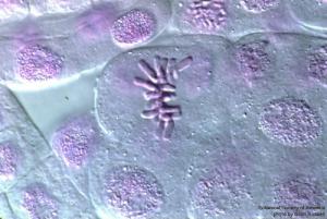
Metaphase. Chromosomes are aligned on the metaphase plate
|
copyright: Scott Russell, BSA
license: http://images.botany.org/index.html#license |
Image
|
Mitosis

|

|
|
15-003h
|
|
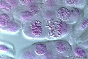
Anaphase A. Chromosomes separate, migrating from the metaphase plate to the poles of the cell
|
copyright: Scott Russell, BSA
license: http://images.botany.org/index.html#license |
Image
|
Mitosis

|

|
|
15-004h
|
|
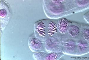
Anaphase B. Chromosomes have separated. In Anaphase B, the spindle poles separate and become more distant
|
copyright: Scott Russell, BSA
license: http://images.botany.org/index.html#license |
Image
|
Mitosis

|

|
|
15-005h
|
|
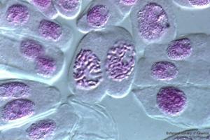
Telophase. Nuclear division is complete, awaiting cytokinesis
|
copyright: Scott Russell, BSA
license: http://images.botany.org/index.html#license |
Image
|
Mitosis

|

|
|
15-006h
|
|
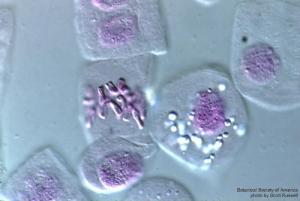
Two cells: Late-metaphase cell on the left. A gravitropic sensing root cell with statocytes on the right
|
copyright: Scott Russell, BSA
license: http://images.botany.org/index.html#license |
Image
|
Mitosis

|

|