|
13-075h
|
|
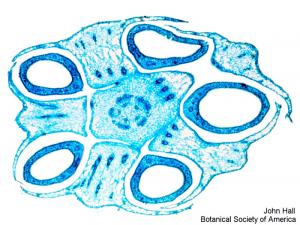
Aquilegia, x.s.
|
copyright: John Hall, BSA
license: http://images.botany.org/index.html#license |
Image
|
Plant Anatomy

|

|
|
13-076h
|
|
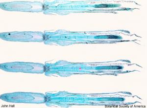
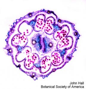
Helianthus florets, l.s. epigynous
|
copyright: John Hall, BSA
license: http://images.botany.org/index.html#license |
Image
|
Plant Anatomy
|
|
|
13-077h
|
|
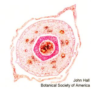
Helianthus, x.s., base of nectary
|
copyright: John Hall, BSA
license: http://images.botany.org/index.html#license |
Image
|
Plant Anatomy
|
|
|
13-078h
|
|
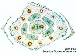
Helianthus, x.s., free filaments
|
copyright: John Hall, BSA
license: http://images.botany.org/index.html#license |
Image
|
Plant Anatomy
|
|
|
13-079h
|
|
Helianthus, x.s., fused anthers
|
copyright: John Hall, BSA
license: http://images.botany.org/index.html#license |
Image
|
Plant Anatomy
|
|
|
13-080h
|
|
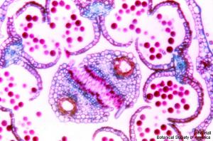
Enlargement of fused anthers
|
copyright: John Hall, BSA
license: http://images.botany.org/index.html#license |
Image
|
Plant Anatomy
|
|
|
13-081h
|
|
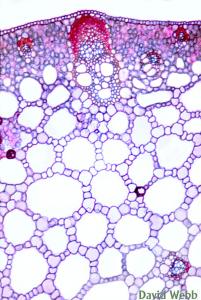
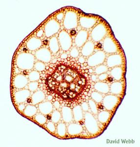
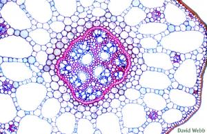
Potamogeton stem, cross section
|
copyright: John Hall, BSA
license: http://images.botany.org/index.html#license |
Image
|
Plant Anatomy
|
|
|
13-082h
|
|
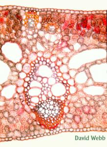
Potamogeton stem, cross section
|
copyright: John Hall, BSA
license: http://images.botany.org/index.html#license |
Image
|
Plant Anatomy
|
|
|
13-083h
|
|
Potamogeton stem, cross section
|
copyright: John Hall, BSA
license: http://images.botany.org/index.html#license |
Image
|
Plant Anatomy
|
|
|
13-084h
|
|
Acorus (sweet flag) leaf, cross section
|
copyright: John Hall, BSA
license: http://images.botany.org/index.html#license |
Image
|
Plant Anatomy
|
|
|
14-001h
|
|
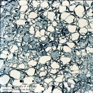
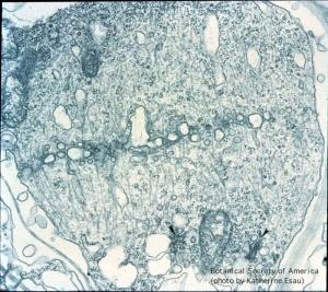
Young parenchyma tissue cut parallel with the epidermis of Euphorbia pulcherrima (poinsettia). Plasmodesmata present in cell walls remain united between cells during intercellular spaces development.
|
copyright: Katherine Esau, BSA
license: http://images.botany.org/index.html#license |
Image
|
Cellular Communication Channels
|
|
|
14-002h
|
|
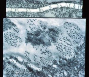
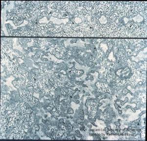
Parenchyma cells in phloem of Cucurbita maxima leaf. In one view, plasmodesmata are shown in longitudinal views, and in transverse views in primary pit fields.
|
copyright: Katherine Esau, BSA
license: http://images.botany.org/index.html#license |
Image
|
Cellular Communication Channels
|
|
|
14-003h
|
|
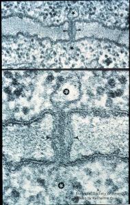
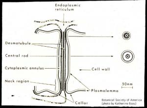
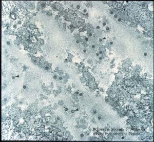
Plasmodesmatal structure in longitudinal section of Phaseolus vulgaris root tip. Cell walls are lined with plasmadesmata between contiguous cells. Demotubule extends to the lumina of ER cisternae.
|
copyright: Katherine Esau, BSA
license: http://images.botany.org/index.html#license |
Image
|
Cellular Communication Channels
|
|
|
14-004h
|
|
Constrictions at both ends of plasmodesma presumably have valve-like function.
|
copyright: Katherine Esau, BSA
license: http://images.botany.org/index.html#license |
Image
|
Cellular Communication Channels
|
|
|
14-005h
|
|
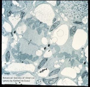
Plasmodesmata originating during cell division. Mitosis associated with accumulation of vesicles in the equatorial plane. This is first stage in cell-wall formation of Nicotiana tabacum mesophyll.
|
copyright: Katherine Esau, BSA
license: http://images.botany.org/index.html#license |
Image
|
Cellular Communication Channels
|
|
|
14-006h
|
|
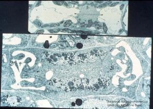
Mitosis in longitudinal divisions of elongated cells. Earlier stage in Nicotiana tabacum; later stage (more extended cell plate) in Sonchus oleraceus.
|
copyright: Katherine Esau, BSA
license: http://images.botany.org/index.html#license |
Image
|
Cellular Communication Channels
|
|
|
14-007h
|
|
Late telophase (nuclei not shown). Microtubules assemble into a phragmoplast centrifugally around the cell plate toward the lateral mother-cell walls (petiole of Echium judaeum).
|
copyright: Katherine Esau, BSA
license: http://images.botany.org/index.html#license |
Image
|
Cellular Communication Channels
|
|
|
14-008h
|
|
Longitudinal and partial surface views of growing cell plates showing involvement of ER in formation of plasmodesmata. ER tubules may be included in openings in the cell plate. Phaseolus vulgaris root tip.
|
copyright: Katherine Esau, BSA
license: http://images.botany.org/index.html#license |
Image
|
Cellular Communication Channels
|
|
|
14-009h
|
|
Cell plate has become the cell wall with fully formed plasmodesmata. Incomplete section made parallel with the surface of the cell wall. Phaseolus vulgaris root tip.
|
copyright: Katherine Esau, BSA
license: http://images.botany.org/index.html#license |
Image
|
Cellular Communication Channels
|
|
|
14-010h
|
|
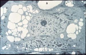
Young sieve element and companion cell, the latter narrow, with a narrow nucleus. Cell wall between the two cells and plasmodesmata in future sieve plates. (Nicotiana tabacum root tip)
|
copyright: Katherine Esau, BSA
license: http://images.botany.org/index.html#license |
Image
|
Cellular Communication Channels
|
|
