|
35-198
|
|
|
copyright: Ronald L. Stuckey, BSA
license: http://images.botany.org/index.html#license |
Image
|
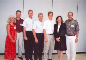
BSA Members

|

|
|
35-199
|
|
|
copyright: Ronald L. Stuckey, BSA
license: http://images.botany.org/index.html#license |
Image
|
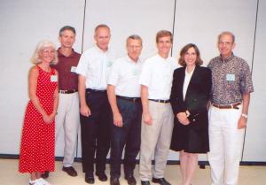
BSA Members
|
|
|
35-200
|
|
|
copyright: Ronald L. Stuckey, BSA
license: http://images.botany.org/index.html#license |
Image
|
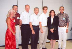
BSA Members
|
|
|
35-201
|
|
|
copyright: Ronald L. Stuckey, BSA
license: http://images.botany.org/index.html#license |
Image
|
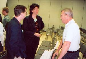
BSA Members
|
|
|
35-202
|
|
|
copyright: Ronald L. Stuckey, BSA
license: http://images.botany.org/index.html#license |
Image
|
BSA Members
|
|
|
35-203
|
|
|
copyright: Ronald L. Stuckey, BSA
license: http://images.botany.org/index.html#license |
Image
|
BSA Members
|
|
|
35-204
|
|
|
copyright: Ronald L. Stuckey, BSA
license: http://images.botany.org/index.html#license |
Image
|
BSA Members
|
|
|
35-205
|
|
|
copyright: Ronald L. Stuckey, BSA
license: http://images.botany.org/index.html#license |
Image
|
BSA Members
|
|
|
35-206
|
|
|
copyright: Ronald L. Stuckey, BSA
license: http://images.botany.org/index.html#license |
Image
|
BSA Members
|
|
|
35-207
|
|
|
copyright: Ronald L. Stuckey, BSA
license: http://images.botany.org/index.html#license |
Image
|
BSA Members
|
|
|
35-208
|
|
|
copyright: Ronald L. Stuckey, BSA
license: http://images.botany.org/index.html#license |
Image
|
BSA Members
|
|
|
aajb2-001
|
|
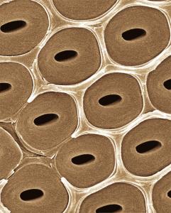
Alternate intervessel pits showing pit borders and pit apertures in the wood of Petersianthus macrocarpus (Lecythidaceae). Lens et al. report that several wood features, including among others vessel pits, type of vessel perforation and the structure of the rays, are phylogenetically informative at the subfamily level. See "A search for phylogenetically informative wood characters within Lecythidaceae s.l." American Journal of Botany 94(4), pp. 483-502.
|
copyright: -
license: - |
Image
|
Plant Anatomy
|
|
|
abot79-1
|
|
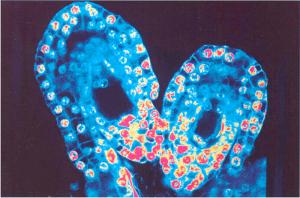
Longitudinal sections of two ovules of the orchid, Epipactis helleborina, photograhped from an image on the confocal laser scanning microscope and artificially colorized.
Article Title: The Development of the Female Gametophyte of Epipactis (Orchidaceae) and its Inference for Reproductive Ecology
Abstract: The development of the female gametophytes of Epipactis atrorubens, E. helleborine, and E. palustris have been investigated using confocal scanning laser microscopy. These species have T-tetrads and eight-nucleate female gametophytes in common. The development of the female gametophyte is monosporic in accordance with the Polygonum type. Furthermore, the outer integument grows slowly and reaches to the middle of the inner integument, when the female gametophyte is mature. In the first two species, T-tetrads develop prior to anthesis, which may be correlated with autogamy. The development of the female gametophyte of E. palustris appears to be similar in plants from different localities.
|
copyright: Margit Fredrikson, BSA
license: http://images.botany.org/index.html#license |
Image
|
Structure and Development
|
|
|
abot79-10
|
|
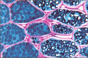
Homogeneous (all blue) and heterogeneous (with white globoids) protein bodies occur in different cells in the cotyledon of Lupinus luteus.
|
copyright: Rehana Asghar, BSA
license: http://images.botany.org/index.html#license |
Image
|
Structure and Development
|
|
|
abot79-11
|
|
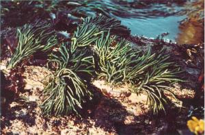
Codium fragile ("dead man's fingers"), a siphonous green alga, along the coast of Santa Cruz County, California.
|
copyright: Lynda J. Goff, BSA
license: http://images.botany.org/index.html#license |
Image
|
Systematics and Ecology
|
|
|
abot79-12
|
|
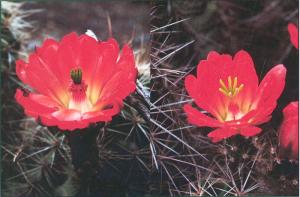
(Left) functional male flower of Echinocereus coccineus (Cactaceae) showing pollen-filled anthers surrounding the base of the stigma lobes. (Right) Functional felmale flower of E. coccineus from a different plant showing reduced filaments and empty anther sacs held below the stigma lobes.
|
copyright: M. Timm Hoffman, BSA
license: http://images.botany.org/index.html#license |
Image
|
Reproductive Biology
|
|
|
abot79-2
|
|
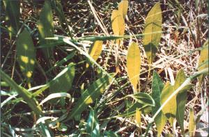
Seedlings of the compass plant, Silphium laciniatum, in a restored prairie on the Iowa State University campus, Ames, Iowa. All seedling leaves are facing the same direction (east).
|
copyright: John M. Pleasants, BSA
license: http://images.botany.org/index.html#license |
Image
|
Reproductive Biology
|
|
|
abot79-3
|
|
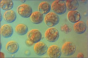
Microspores of Hemerocallis Fulva, isolated from anthers and converted to naked protoplasts by enzymatic treatment that removed the exine and entine (Nomarski interference contrast micography)
Article Title: Experimental Plant Reproductive Biology and Reproductive Cell Manipulation in Higher Plants: Now and the Future
Abstract: Experimental plant reproductive biology has recently emerged as a new form of experimental embryology characterized by the direct experimental isolation and manipulation of reproductive cells and protoplasts. This work is fostered by current multidisciplinary trends in the sciences and serves to deepen our knowledge about the control of reproductive processes by providing novel in vitro material. The techniques of experimental plant reproductive biology also show great potential in providing new means for biotechnology, leading to significant refinements in plant breeding in higher plants that may eventually permit direct reproductive cell engineering. Recent achievements by our research group in experimental manipulation and biological studies of pollen protoplasts, generative cells, sperm cells, and embryo sacs are reviewed, and some ideas about future developments in this new area are presented.
|
copyright: Hong-Yuan Yang, BSA
license: http://images.botany.org/index.html#license |
Image
|
Special Invited Paper
|
|
|
abot79-4
|
|
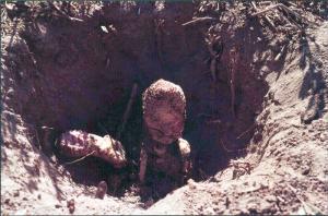
Two plants of Ombrophytum subterraneum (Balanophoraceae), parasitizing Tessaria roots. The plants are reportedly entirely subterranean, even during flowering and seed dispersal; excavation is necessary to locate and study them.
|
copyright: James D. Mauseth, Gloria Montenegro, BSA
license: http://images.botany.org/index.html#license |
Image
|
Structure and Development
|
|
|
abot79-5
|
|
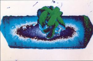
Joined calcium crystals (color transform of scanning electron micrograph) isolated from Capsicum annuum anther connective tissue. These crystals (large one a prism, small one a spherulite), form either together or separately in connective cells during microsporogenesis.
|
copyright: Harry T. Horner, Bruce L. Wagner, BSA
license: http://images.botany.org/index.html#license |
Image
|
Structure and Development
|
|
