|
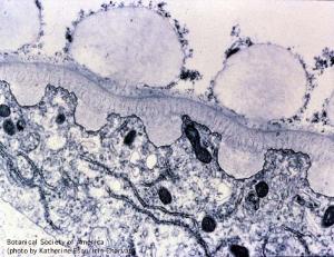
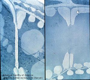
03-017h
|
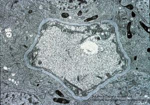
|
Nicotiana tabacum, root
|
copyright: Katherine Esau and Jennifer Thorsch, BSA
license: http://images.botany.org/index.html#license |
Image
|
Phloem Development

|

|
|
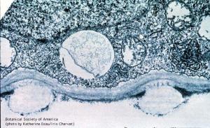
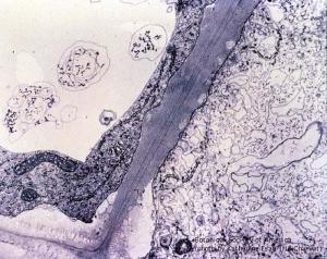
03-018h
|
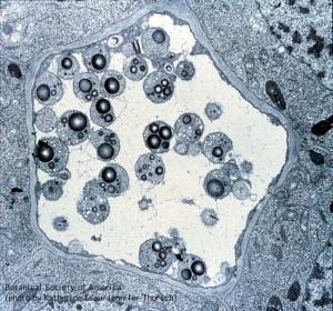
|
Nicotiana tabacum, root
|
copyright: Katherine Esau and Jennifer Thorsch, BSA
license: http://images.botany.org/index.html#license |
Image
|
Phloem Development
|
|
|
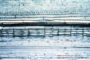
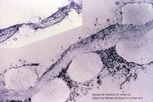
03-019h
|
|
Gossypium hirsutum, petiole
|
copyright: Katherine Esau and Jennifer Thorsch, BSA
license: http://images.botany.org/index.html#license |
Image
|
Phloem Development
|
|
|
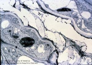
03-020h
|
|
Phaseolus vulgaris, stem
|
copyright: Katherine Esau and Jennifer Thorsch, BSA
license: http://images.botany.org/index.html#license |
Image
|
Phloem Development
|
|
|
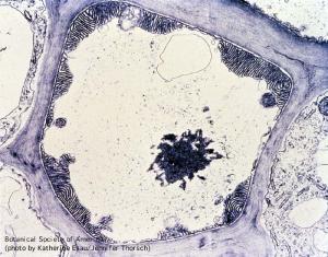
03-021h
|
|
Phaseolus vulgaris, root
|
copyright: Katherine Esau and Jennifer Thorsch, BSA
license: http://images.botany.org/index.html#license |
Image
|
Phloem Development
|
|
|
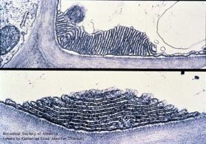
03-022h
|
|
Gossypium hirsutum, stem
|
copyright: Katherine Esau and Jennifer Thorsch, BSA
license: http://images.botany.org/index.html#license |
Image
|
Phloem Development
|
|
|
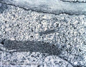
03-023h
|
|
Gossypium hirsutum, petiole
|
copyright: Katherine Esau and Jennifer Thorsch, BSA
license: http://images.botany.org/index.html#license |
Image
|
Phloem Development
|
|
|
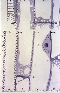
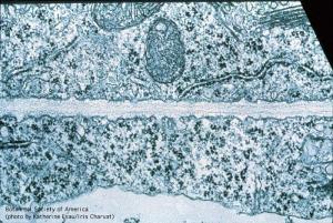
04-001h
|
|
Section through xylem region of a vascular bundle drawn from electron micrographs. Parenchyma cells present; all other cells are tracheary elements in various developmental stages. Scale line, 10 um.
|
copyright: Katherine Esau and Jennifer Thorsch, BSA
license: http://images.botany.org/index.html#license |
Image
|
Xylem Development
|
|
|
04-002h
|
|
Ontogenetic sequence of primary xylem elements (from bottom to top) with annular secondary wall thickenings (rings) gradating to helical thickenings in a moderately stretched cell.
|
copyright: Katherine Esau and Jennifer Thorsch, BSA
license: http://images.botany.org/index.html#license |
Image
|
Xylem Development
|
|
|
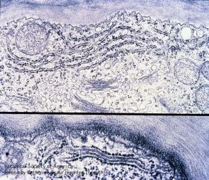
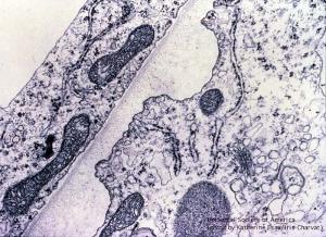
04-003h
|
|
Side wall between two differentiating vessel members. Fibrous material is present in vesicles connected with the near wall. Middle lamella and connections between microtubules and plasmalemma.
|
copyright: Katherine Esau and Jennifer Thorsch, BSA
license: http://images.botany.org/index.html#license |
Image
|
Xylem Development
|
|
|
04-004h
|
|
Side wall between differentiating vessel members. Secondary wall deposition not yet evident. In the older cell (below), microtubules concentrate near young helical thickenings and are absent along the primary wall.
|
copyright: Katherine Esau and Jennifer Thorsch, BSA
license: http://images.botany.org/index.html#license |
Image
|
Xylem Development
|
|
|
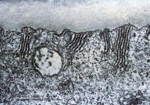
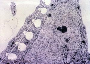
04-005h
|
|
Side wall between vessel members with differentiating helical thickenings. More mature cell (right) indicated by larger amount of secondary wall deposition. Vesicles between plasmalemma and secondary wall are evident.
|
copyright: Katherine Esau and Jennifer Thorsch, BSA
license: http://images.botany.org/index.html#license |
Image
|
Xylem Development
|
|
|
04-006h
|
|
Wall between older (right) and younger (left) vessel members. Microtubules accumulate near the secondary wall in younger cell. Membrane-bound vesicles appear as blebs on plasmalemma. Primary wall of older cell are partly hydrolyzed.
|
copyright: Katherine Esau and Jennifer Thorsch, BSA
license: http://images.botany.org/index.html#license |
Image
|
Xylem Development
|
|
|
04-007h
|
|
Vessel member above, parenchyma cell below. Primary wall is partly hydrolyzed in the vessel member. Protoplasmic residue covers the secondary and hydrolyzed primary wall.
|
copyright: Katherine Esau and Jennifer Thorsch, BSA
license: http://images.botany.org/index.html#license |
Image
|
Xylem Development
|
|
|
04-008h
|
|
Degradation of primary wall of vessenl member is almost complete. Primary wall of other cell is partly hydrolyzed between the coils of the helix. Older helix subjected to greater amount of stretching.
|
copyright: Katherine Esau and Jennifer Thorsch, BSA
license: http://images.botany.org/index.html#license |
Image
|
Xylem Development
|
|
|
04-009h
|
|
Later stage of end wall degradation.
|
copyright: Katherine Esau and Jennifer Thorsch, BSA
license: http://images.botany.org/index.html#license |
Image
|
Xylem Development
|
|
|
04-010h
|
|
Rim of secondary wall material (top) has been deposited around the margin of the end wall delimiting the future perforation. Part of the end wall not covered by the rim (bottom) has been removed.
|
copyright: Katherine Esau and Jennifer Thorsch, BSA
license: http://images.botany.org/index.html#license |
Image
|
Xylem Development
|
|
|
04-011h
|
|
Vesicles occur near the surface of the exposed wall part and some also within the immature rim. Microtubules near the rim at arrowheads.
|
copyright: Katherine Esau and Jennifer Thorsch, BSA
license: http://images.botany.org/index.html#license |
Image
|
Xylem Development
|
|
|
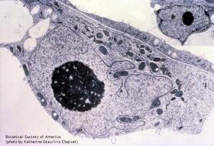
04-012h
|
|
Nucleus in a xylem parenchyma cell. At cw, primary cell wall between two parenchyma cells.
|
copyright: Katherine Esau and Jennifer Thorsch, BSA
license: http://images.botany.org/index.html#license |
Image
|
Xylem Development
|
|
|
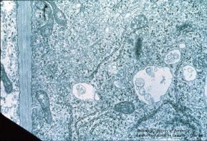
04-013h
|
|
Protoplast of differentiating vessel member with part of end wall. Details in the protoplast include dictyosome with vesicles, mitochondria, microbodies, rough and smooth ER, and autophagic vacuoles.
|
copyright: Katherine Esau and Jennifer Thorsch, BSA
license: http://images.botany.org/index.html#license |
Image
|
Xylem Development
|
|
